Sleep & Wellness
You show up at a sleep center at nine at night, and by the time you’re tucked into bed you’re wearing more wires than a recording studio. It looks intimidating, and it’s fair to wonder what all of that equipment is actually measuring, and whether it’s even possible to fall asleep with electrodes glued to your scalp and a clip on your finger. The honest answer is that a sleep study, formally called polysomnography, is one of the most information-dense medical tests you’ll ever take: in a single night it records your brain activity, your eye movements, your muscle tone, your heartbeat, your breathing, and your blood oxygen levels, all stitched together on a single timeline so a sleep physician can see exactly what your body did, minute by minute, while you were unconscious. This guide walks through every signal a sleep study actually captures, what those signals are used to diagnose, how the different types of sleep studies compare, and how to make sense of the numbers that show up on your results.
Quick answer: what does a sleep study test for?
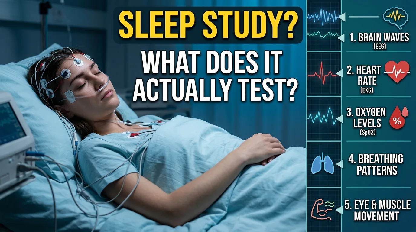
A standard sleep study (polysomnography) simultaneously records brain waves (EEG), eye movements (EOG), chin and leg muscle activity (EMG), heart rhythm (ECG), airflow through your nose and mouth, chest and belly breathing effort, blood oxygen saturation, and snoring, body position, and behavior via audio and video. Together, these signals let a sleep physician identify what stage of sleep you’re in at any given moment and flag abnormal events, pauses in breathing, oxygen drops, abnormal limb movements, or unusual brain activity, that point toward conditions like obstructive sleep apnea, central sleep apnea, narcolepsy, periodic limb movement disorder, REM sleep behavior disorder, or nocturnal seizures. It’s considered the gold-standard test for diagnosing sleep-related breathing disorders because no other single test captures this many overlapping systems at once.
Every signal a sleep study records, and what each one is for
A polysomnogram typically involves somewhere between 15 and 25 individual sensors, and every one of them exists to answer a specific clinical question. Here’s what’s actually being measured, channel by channel.

Brain waves (EEG)
Electrodes attached to your scalp, usually at the frontal, central, and occipital positions on both sides of your head, record your brain’s electrical activity throughout the night. This is the single most important signal for determining sleep architecture: it’s how a technologist can tell whether you’re awake, in light sleep, in deep slow-wave sleep, or in REM sleep at any given 30-second window. Distinct brain-wave patterns also reveal arousals, brief, often unnoticed awakenings that fragment sleep without you remembering them the next morning, and can flag unusual electrical activity consistent with nocturnal seizures.
Eye movements (EOG)
Small electrodes placed near the outer corners of each eye track eye movement. This channel exists almost entirely to help identify REM sleep, the stage defined by rapid, darting eye movements (and the stage most associated with vivid dreaming). Slow, rolling eye movements are also one of the earliest signs that you’re drifting from wakefulness into light sleep.
Muscle activity (EMG)
A small sensor under your chin measures muscle tone there, which normally drops sharply during REM sleep as your body enters a near-paralysis state that stops you from physically acting out dreams. If chin muscle tone doesn’t drop the way it should during REM, that’s one of the hallmark findings in REM sleep behavior disorder. Separate sensors on your shins record leg muscle activity, which is used to detect periodic limb movements, repetitive, involuntary jerks that can fragment sleep without you being fully aware of them.
Heart rhythm (ECG)
A simplified electrocardiogram tracks your heart rate and rhythm throughout the night. Sleep-disordered breathing places real strain on the cardiovascular system, and it’s common to see heart rate slow down during an apnea event and then spike as breathing resumes, a pattern that helps confirm a respiratory event was severe enough to trigger a stress response. This channel can also catch nighttime arrhythmias that a patient might otherwise never know about.
Airflow and breathing effort
This is arguably the centerpiece of a diagnostic sleep study. A nasal pressure sensor and an oral-nasal thermal airflow sensor sit just under your nose to measure the air actually moving in and out of your airway, while elastic belts around your chest and abdomen measure the physical effort your body is making to breathe. Comparing these two signals is how technologists tell the difference between an obstructive event (your airway is blocked, so your chest and belly are working hard but air still isn’t getting through) and a central event (your brain briefly stops sending the signal to breathe at all, so there’s no effort and no airflow).
Blood oxygen saturation (pulse oximetry)
A small clip, usually on a finger, uses light absorption to continuously estimate the percentage of oxygen carried in your blood. Healthy oxygen saturation generally stays above roughly 95%; when breathing is repeatedly interrupted, saturation can drop noticeably with each event, and the frequency and depth of these drops (sometimes summarized separately as the Oxygen Desaturation Index, or ODI) is one of the clearest signs of how physiologically significant a breathing disruption really is.
Snoring, body position, and video
A small microphone tracks snoring intensity and pattern, sensors detect whether you’re lying on your back, side, or stomach (since sleep apnea is frequently worse when sleeping on your back), and infrared video with audio lets a technologist, and later, the interpreting physician, review anything unusual: sleepwalking, unusual movements, or confirming that a reported “gasping” episode lines up with what the other channels recorded.
The four types of sleep studies, and why the type matters
Not every “sleep study” records all of the above. Sleep medicine classifies testing into four types based on how many channels are recorded and where the test happens, and knowing the difference matters because it affects what the test can and can’t reliably diagnose.
| Type | What it records | Where it happens | Best for |
|---|---|---|---|
| Type I | Full polysomnography – all EEG, EOG, EMG, ECG, airflow, effort, and oxygen channels | In-lab, technologist present overnight | Comprehensive diagnosis: sleep apnea, narcolepsy, limb movement disorders, parasomnias, seizures |
| Type II | The same full channel set as Type I, unattended | Typically at home | Full diagnostic detail without an overnight lab stay (less common due to setup complexity) |
| Type III | Limited channels – usually airflow, respiratory effort, oxygen saturation, and heart rate, no EEG | At home | Home sleep apnea testing (HSAT) for patients with a high pre-test likelihood of moderate-to-severe obstructive sleep apnea and no other suspected sleep disorder |
| Type IV | One or two channels, often just oxygen saturation and airflow or a wearable-based signal | At home | Basic apnea screening; least detailed, higher chance of under-diagnosing mild or borderline cases |
The practical takeaway: because home sleep apnea tests (Type III and IV) don’t record brain waves, they can’t tell you what stage of sleep you were in, can’t detect arousals that don’t affect breathing, and can’t diagnose narcolepsy, periodic limb movement disorder, REM sleep behavior disorder, or nocturnal seizures. They’re a reasonable, more affordable, and more convenient option specifically for confirming moderate-to-severe obstructive sleep apnea in the right patient, but they’re not a substitute for a full in-lab study when the clinical picture is more complicated, for example, if you have a lot of daytime sleepiness but a home test comes back negative, or if a doctor suspects something beyond a straightforward breathing disorder.
What conditions a sleep study can actually diagnose

Because polysomnography records so many overlapping body systems at once, it’s used well beyond just confirming snoring. Conditions a full, in-lab sleep study is used to diagnose or evaluate include:
- Obstructive sleep apnea (OSA) – repeated partial or complete airway collapse during sleep, identified by effort continuing while airflow drops or stops.
- Central sleep apnea – breathing pauses where the brain temporarily stops signaling the breathing muscles at all, identified by both effort and airflow dropping together.
- Narcolepsy – usually confirmed with an overnight polysomnogram followed the next day by a Multiple Sleep Latency Test (MSLT), which measures how quickly you fall asleep during a series of scheduled daytime naps.
- Periodic limb movement disorder – repetitive leg jerks during sleep, picked up by the leg EMG channels, that are frequent and disruptive enough to fragment sleep.
- REM sleep behavior disorder – a failure of the normal muscle paralysis that should occur during REM sleep, which can cause people to physically act out dreams.
- Nocturnal seizures – unusual, seizure-consistent electrical activity on the EEG channel during sleep.
- Complex or treatment-related insomnia – when insomnia is suspected to overlap with an underlying physiological sleep disorder rather than being purely behavioral.
If you’re trying to figure out whether your own symptoms. Loud snoring, gasping awake, excessive daytime sleepiness, point toward sleep apnea specifically, our guide on the warning signs of sleep apnea you shouldn’t ignore breaks down what to watch for before you even get to the testing stage.
How the results are scored: AHI, RDI, and sleep efficiency
Once the raw data is collected, a certified sleep technologist scores the recording in 30-second segments, and a sleep physician reviews and interprets it. A few key numbers show up on almost every report.
Apnea-Hypopnea Index (AHI)
The AHI is the number of apneas (near-complete pauses in breathing lasting at least 10 seconds) plus hypopneas (partial reductions in airflow of roughly 30% or more, lasting at least 10 seconds and accompanied by an oxygen drop or an arousal) averaged per hour of sleep. Under American Academy of Sleep Medicine (AASM) adult criteria, this is generally interpreted as:
- Normal: fewer than 5 events per hour
- Mild: 5 to 14 events per hour
- Moderate: 15 to 29 events per hour
- Severe: 30 or more events per hour
Children are scored against much stricter thresholds, since even a small number of breathing disruptions is considered clinically significant at a young age.
Oxygen Desaturation Index (ODI) and other supporting numbers
The ODI captures how often, and how far, blood oxygen levels drop during the night, which helps a physician judge how physiologically stressful the breathing events actually were; two people can have a similar AHI but very different oxygen profiles. Reports also typically include total sleep time, sleep efficiency (the percentage of time in bed actually spent asleep), time spent in each sleep stage, and the arousal index, which reflects how fragmented your sleep was overall, independent of breathing.
Sleep physicians are generally taught not to rely on AHI alone. A patient with a technically “mild” AHI but severe oxygen drops or significant daytime symptoms may still warrant treatment, while borderline numbers are always interpreted alongside the patient’s actual symptoms and health history, which is exactly why a physician, not just a number on a printout, makes the final diagnosis.
Will a sleep study actually work if I can’t sleep normally there?
This is one of the most common worries, and it’s a fair one; sleeping in an unfamiliar room with wires attached to your body is not most people’s idea of a normal night. Sleep researchers even have a name for the well-documented pattern of worse-than-usual sleep on the first night in a new environment: the “first-night effect.” The reassuring part is that a diagnostic sleep study doesn’t need you to sleep perfectly to be useful. Technologists only need a few hours of scoreable sleep, across multiple stages, to identify a clear pattern of disordered breathing or abnormal movement, and conditions like obstructive sleep apnea tend to show up reliably even on a night of lighter, more fragmented sleep than usual. If you want a full walkthrough of what an overnight visit actually looks like from arrival to wake-up, see our guide on what happens during a nighttime sleep study.
Common misconceptions about what a sleep study tests for
- “It only checks for sleep apnea.” A full in-lab study evaluates brain activity, muscle activity, and heart rhythm too, which is why it can catch narcolepsy, limb movement disorders, parasomnias, and nocturnal seizures, not just breathing problems.
- “A home test is just as thorough as an in-lab test.” Home sleep apnea tests are validated for a specific purpose, confirming likely moderate-to-severe OSA in an otherwise straightforward case, but they skip brain-wave monitoring entirely, so they can miss other conditions or underestimate severity in more complex patients.
- “One bad number means I definitely have a disorder.” A single elevated AHI or a rough night’s sleep during testing is interpreted in context by a physician, alongside your symptoms and history, not read in isolation.
- “The wires will keep me awake all night, so the test won’t be accurate.” Modern sensors are lightweight and the wiring is bundled to allow normal movement; as above, the test is designed to extract a diagnosis from a realistically imperfect night of sleep.
- “Sleep studies are only for people who snore loudly.” Excessive daytime sleepiness, unexplained morning headaches, unrefreshing sleep, and witnessed breathing pauses are all valid reasons a doctor might order testing, independent of snoring.
Frequently asked questions
Does a sleep study test for anything besides sleep apnea?
Yes. Because a full polysomnogram records brain waves, eye movements, and muscle activity in addition to breathing, it’s also used to help diagnose narcolepsy (typically paired with a next-day Multiple Sleep Latency Test), periodic limb movement disorder, REM sleep behavior disorder, and nocturnal seizure activity. Home sleep apnea tests, by contrast, are narrower and are validated specifically for detecting moderate-to-severe obstructive sleep apnea.
How accurate are home sleep apnea tests compared to in-lab studies?
Home sleep apnea tests (Type III devices) are considered reasonably accurate for confirming moderate-to-severe obstructive sleep apnea in patients who have a high likelihood of the condition based on symptoms and risk factors, and they’re widely used for that reason. However, because they don’t record EEG, they can’t measure actual sleep time or stage; the “hours” used in their calculations are usually total recording time, not confirmed sleep time, which means they can underestimate severity, especially in people with fragmented sleep or a lower overall likelihood of OSA. An in-lab study remains the more reliable choice when the diagnosis is unclear or another disorder is suspected.
Can a sleep study diagnose insomnia?
Insomnia itself is usually diagnosed based on your reported symptoms and sleep history rather than a sleep study, since the core problem, difficulty falling or staying asleep, is something you experience rather than something a machine needs to confirm. A sleep study is more often ordered for someone with insomnia symptoms when a doctor suspects an underlying physiological cause, such as undiagnosed sleep apnea or periodic limb movements, might be contributing to or masquerading as insomnia.
Do sleep studies check oxygen levels?
Yes, continuous pulse oximetry is a standard part of every level of sleep study, from a full in-lab polysomnogram down to a basic home screening device. Oxygen saturation trends and the frequency of desaturation events are among the most clinically important pieces of data a sleep study produces, because they show how much a breathing disruption is actually affecting the body, not just how often it happens.
What happens if my sleep study results come back abnormal?
An abnormal result, commonly an elevated AHI consistent with sleep apnea, typically leads to a follow-up conversation with your ordering physician or a board-certified sleep medicine specialist about treatment options. For obstructive sleep apnea, that often means a CPAP titration study or an auto-adjusting CPAP trial, oral appliance therapy, or, in select cases, evaluation for surgery; for other conditions identified on the study, treatment is tailored to that specific diagnosis. The report itself is a starting point for a conversation, not a treatment plan on its own.
Will I actually fall asleep during a sleep study?
Most people do, even though it usually takes longer than at home and the sleep tends to be lighter and more fragmented than usual, the well-documented “first-night effect.” Sleep centers are designed to make this easier, with private rooms, comfortable bedding, and dim lighting, and technologists generally only need a handful of hours of scoreable sleep across different stages to gather enough data for an accurate diagnosis. If you’re worried about this specifically, our guide on what to expect during your first sleep study covers practical tips for settling in.
One last note worth repeating: everything above describes what a sleep study measures and how it’s generally interpreted, not a diagnosis for any individual case. If you’re experiencing symptoms like loud snoring, gasping awake, or persistent daytime fatigue, the right next step is talking to a doctor or an accredited sleep center, who can determine whether testing is appropriate and which type of study fits your situation.




Leave a Reply