Sleep & Wellness
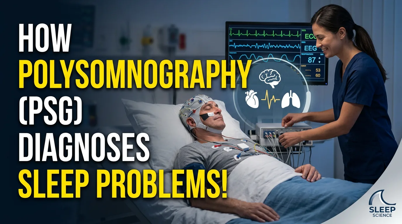
Polysomnography, the technical name for an overnight sleep study, is often described as the “gold standard” test in sleep medicine, but that phrase doesn’t explain much on its own. What actually makes it the gold standard is the way it combines brain, eye, muscle, heart, and breathing data into a single, minute-by-minute record that a sleep physician can score against established clinical criteria. That combination is what allows one overnight test to distinguish between a dozen different possible explanations for the same complaint, say, daytime exhaustion, instead of just confirming or ruling out one narrow thing. This guide breaks down exactly how a diagnosis gets made from a polysomnogram: how the data is scored, what specific patterns point to which conditions, and what happens between the night of testing and the moment your doctor tells you what’s wrong.
Quick answer: how does polysomnography lead to a diagnosis?
Polysomnography diagnoses sleep problems by recording brain waves, eye movements, muscle activity, heart rhythm, breathing effort, airflow, and blood oxygen simultaneously overnight, then having a certified technologist score the recording in 30-second segments and a board-certified sleep physician interpret the scored data against established diagnostic criteria. Specific patterns in that data, repeated drops in airflow with continued breathing effort, abnormal muscle tone during REM sleep, excessive leg movements, or unusual brain activity, each point toward a specific diagnosis, such as obstructive sleep apnea, REM sleep behavior disorder, periodic limb movement disorder, or nocturnal seizures. The physician then combines those findings with your reported symptoms and medical history to reach a final diagnosis and recommend treatment.
From raw signals to a diagnosis: how scoring actually works
A single night of polysomnography generates an enormous amount of raw data, often eight or more distinct signal channels recorded continuously for six to eight hours. Turning that into something a doctor can act on happens in two stages.

Step one: technologist scoring
A registered polysomnographic technologist reviews the entire recording, typically in 30-second windows called “epochs,” and manually (with software assistance) labels what’s happening in each one: which sleep stage you’re in, whether an apnea or hypopnea occurred, whether there was a leg movement, an arousal, or an abnormal EEG pattern. This scoring follows standardized rules published by the American Academy of Sleep Medicine (AASM), which is what makes results comparable across different labs and technologists; an apnea scored in one sleep lab should meet the same definition as an apnea scored in another.
Step two: physician interpretation
A board-certified sleep medicine physician then reviews the scored data as a whole, not just the summary numbers. This matters because two people can have a similar Apnea-Hypopnea Index (AHI) but very different clinical pictures; one might have brief, shallow events with minimal oxygen impact, while another has fewer but much more severe events with significant oxygen drops and cardiac strain. The physician weighs the full picture, including your symptoms, health history, and the raw data patterns, before issuing a formal diagnosis. This is also why automated, software-generated scoring on home sleep apnea devices is generally expected to be reviewed by a physician before being used for diagnosis, rather than accepted at face value.
How polysomnography diagnoses obstructive and central sleep apnea
Sleep apnea is the condition polysomnography is most associated with, and the diagnostic logic is a good example of how the different channels work together.
Telling obstructive from central events
The airflow sensors (nasal pressure and oral-nasal thermal sensors) and the respiratory effort belts around the chest and abdomen work as a pair. In an obstructive apnea, the airway physically collapses or narrows, so airflow drops sharply even though the chest and belly are still visibly straining to breathe. In a central apnea, the brain briefly stops sending the signal to breathe altogether, so both airflow and respiratory effort drop together, with no straining at all. A mixed apnea shows a central pattern at the start of the event that shifts into an obstructive pattern before breathing resumes. This distinction matters clinically because obstructive and central sleep apnea are typically treated differently.
Scoring severity with the Apnea-Hypopnea Index
Every scored apnea (an airflow reduction of about 90% or more for at least 10 seconds) and hypopnea (a partial reduction of roughly 30% or more for at least 10 seconds, paired with either an oxygen desaturation or an arousal) gets counted and averaged per hour of sleep to produce the AHI. Under widely used adult diagnostic thresholds:
- Normal: fewer than 5 events per hour
- Mild OSA: 5–14 events per hour
- Moderate OSA: 15–29 events per hour
- Severe OSA: 30 or more events per hour
Physicians also weigh oxygen desaturation patterns and reported daytime symptoms alongside the raw AHI, since a technically “mild” score paired with significant oxygen drops or severe daytime sleepiness may still warrant treatment. If you’re wondering whether your own symptoms line up with sleep apnea before testing even comes into the picture, our guide on the warning signs of sleep apnea you shouldn’t ignore is a useful starting point.
CPAP titration: diagnosis leading directly into treatment
Once obstructive sleep apnea is confirmed, many patients undergo a second type of polysomnogram called a CPAP titration study, where a technologist gradually adjusts the pressure of a continuous positive airway pressure machine overnight while monitoring the same channels used for diagnosis to find the specific pressure setting that eliminates most breathing events. Some sleep centers combine both steps into a single “split-night” study, diagnosing sleep apnea in the first half of the night and titrating CPAP pressure in the second half, if the apnea is confirmed to be severe enough early on.
How polysomnography diagnoses conditions beyond sleep apnea
Because a full polysomnogram also records brain activity and muscle tone, it’s used to identify several conditions that have nothing directly to do with breathing.
Narcolepsy
Narcolepsy is generally diagnosed using an overnight polysomnogram followed the next day by a Multiple Sleep Latency Test (MSLT), which offers a series of scheduled short nap opportunities and measures both how quickly you fall asleep and whether you enter REM sleep abnormally fast. The overnight polysomnogram first rules out other causes of excessive daytime sleepiness, like undiagnosed sleep apnea, before the MSLT results are interpreted as consistent with narcolepsy. For a closer look at how disruptive this specific condition can be day to day, see our guide on Is Narcolepsy Dangerous? What You Need to Know.
Periodic limb movement disorder and restless leg syndrome
Sensors on the shins record leg muscle activity throughout the night. When a technologist identifies a certain frequency of repetitive, stereotyped leg movements, particularly movements linked to arousals or sleep fragmentation, that pattern supports a diagnosis of periodic limb movement disorder. This is a distinct (though sometimes overlapping) diagnosis from restless leg syndrome, which is a subjective symptom, typically diagnosed by clinical history rather than PSG, describing an uncomfortable urge to move the legs, usually in the evening or at rest; our guide on What Causes Restless Leg Syndrome During Sleep? covers that distinction in more depth.
REM sleep behavior disorder
During normal REM sleep, chin and limb muscles should show a marked drop in tone, effectively holding the body still while the brain is highly active and dreaming. In REM sleep behavior disorder, that muscle paralysis doesn’t fully occur, and the chin EMG channel shows abnormally sustained or excessive muscle activity during REM periods, sometimes correlating with movements or vocalizations captured on the study’s video and audio recording. This is an important diagnosis to get right, since REM sleep behavior disorder has also been linked in research to a higher future risk of certain neurodegenerative conditions, making accurate identification clinically meaningful beyond just the sleep disruption itself. Our guide on Does REM Sleep Behavior Disorder Require Treatment? covers what usually happens after a diagnosis like this.
Nocturnal seizures
Unusual, rhythmic, or otherwise abnormal electrical activity on the EEG channel during sleep can point toward seizure activity that occurs specifically or predominantly at night. Because polysomnography’s EEG montage is less extensive than a dedicated diagnostic EEG, a positive finding here typically leads to a referral for more specialized epilepsy testing rather than a final diagnosis on its own.
Insomnia with a suspected underlying cause
Insomnia itself is usually a clinical diagnosis based on your reported history, not something polysomnography is needed to confirm. But when a doctor suspects that a patient’s insomnia is being driven or worsened by an unrecognized physiological problem, undiagnosed sleep apnea, periodic limb movements, or a circadian rhythm issue, a sleep study can identify that underlying contributor, which changes the treatment approach considerably. If you’re trying to work out whether what you’re dealing with is insomnia at all, our guide on Insomnia vs. Sleep Disturbances: What’s the Difference? is a useful starting point.
What the final report actually contains
A polysomnography report translates the night into a set of standardized measures your doctor will discuss with you, typically including: total sleep time and sleep efficiency (the percentage of time in bed actually asleep), time spent in each sleep stage, the arousal index, the AHI (and often a related Respiratory Disturbance Index that also counts subtler breathing-related arousals), the Oxygen Desaturation Index, lowest recorded oxygen saturation, heart rate trends, and a summary of any leg movements or unusual behavioral or EEG events noted by the technologist. The physician’s written interpretation ties these numbers together into a diagnostic impression and, usually, a treatment recommendation.
In-lab versus home testing: does it change how the diagnosis is made?
It’s worth repeating a point that matters a lot for how a diagnosis gets made: home sleep apnea tests (typically Type III devices) skip the EEG, EOG, and chin EMG channels entirely, recording only airflow, respiratory effort, oxygen saturation, and heart rate. That’s enough to reliably diagnose moderate-to-severe obstructive sleep apnea in a patient who was already a strong clinical candidate for it, but it cannot diagnose narcolepsy, periodic limb movement disorder, REM sleep behavior disorder, or nocturnal seizures, because those diagnoses depend entirely on the brain and muscle channels a home test doesn’t record. If a home test comes back negative despite strong symptoms, or if something beyond straightforward sleep apnea is suspected, an in-lab polysomnogram is the appropriate next step. For a full breakdown of every channel a complete study records and what each one is used for, see our companion guide on what a sleep study actually tests for.
Common misunderstandings about how sleep studies diagnose problems
- “One number tells the whole story.” AHI is important, but physicians weigh it alongside oxygen data, arousal patterns, and your actual symptoms rather than treating it as a single pass/fail score.
- “A normal-looking night means nothing was wrong.” Some conditions, like periodic limb movements or mild upper airway resistance, can be subtle and still clinically meaningful even without dramatic breathing pauses.
- “The test diagnoses you instantly, that night.” Scoring and physician interpretation typically take anywhere from a few days to a couple of weeks after the study before you get formal results.
- “A home test and an in-lab test diagnose the exact same things.” As covered above, home tests are narrower by design and aren’t built to catch non-breathing-related sleep disorders.
A closing note that’s worth taking seriously: everything above explains how polysomnography works and how sleep physicians generally use it, but it isn’t medical advice and it can’t tell you what’s happening in your own body. If you’re experiencing symptoms like loud snoring, witnessed breathing pauses, excessive daytime sleepiness, or unusual nighttime behavior, the right step is to talk to a doctor or an accredited sleep center about whether testing is appropriate for you.
Frequently asked questions
How long does it take to get sleep study results after polysomnography?
It varies by sleep center, but results typically take anywhere from a few days to about two weeks. The recording first has to be manually scored by a certified technologist in 30-second segments, and then a board-certified sleep physician reviews the scored data before writing a formal interpretation that gets sent to your referring doctor.
Can polysomnography diagnose more than one sleep disorder at once?
Yes. Because it records brain waves, eye movements, muscle activity, heart rhythm, and breathing simultaneously, a single overnight study can reveal, for example, both obstructive sleep apnea and periodic limb movement disorder in the same patient, or confirm that a movement disorder rather than a breathing problem is the main driver of someone’s fragmented sleep. This is one of the key advantages of a full in-lab study compared to a narrower home sleep apnea test.
What is the difference between AHI and RDI on a sleep study report?
The Apnea-Hypopnea Index (AHI) counts apneas and hypopneas that meet a specific airflow-reduction threshold, averaged per hour of sleep. The Respiratory Disturbance Index (RDI) typically includes those same events plus “respiratory effort-related arousals”, more subtle breathing disruptions that don’t fully meet the criteria for a hypopnea but still cause a brief arousal from sleep. RDI is generally equal to or slightly higher than AHI, and some sleep physicians consider it a more sensitive measure of upper airway resistance in patients whose symptoms don’t fully match a normal AHI result.
Does a sleep study diagnosis require you to sleep a full eight hours?
No. Sleep technologists generally only need a handful of hours of scoreable sleep across multiple stages, including some REM sleep if possible, to gather enough data for an accurate diagnosis. Many people sleep less than usual during a study due to the unfamiliar environment, a well-documented pattern called the first-night effect, and this rarely prevents a valid diagnosis, particularly for sleep apnea, which tends to show up reliably even during lighter, more fragmented sleep.
Can a sleep study be wrong or miss a diagnosis?
Like any medical test, polysomnography isn’t infallible. Night-to-night variability in sleep apnea severity, an unusually light or restless night, or a condition that happens to not manifest strongly on a given night can all affect results. If symptoms strongly suggest a sleep disorder but a study comes back negative or ambiguous, doctors will sometimes recommend a repeat study or additional testing rather than ruling the condition out based on a single night.
Do you need a referral to get a diagnostic sleep study?
In most healthcare systems, yes, a diagnostic polysomnogram is typically ordered by a physician, often after an initial evaluation of your symptoms, and is billed through insurance as a medical test. This is different from the research-based paid sleep studies some people participate in for compensation rather than diagnosis, which is covered in our guide on paid sleep study opportunities.







Leave a Reply